8th World Congress on
Cancer Research and Oncology
THEME: "Advancing Innovation and Collaboration in Cancer Research for a Healthier Tomorrow"
 23-24 Nov 2026
  Holiday Inn Express Bangkok, Thailand
 THEME: "Advancing Innovation and Collaboration in Cancer Research for a Healthier Tomorrow"
23-24 Nov 2026
Holiday Inn Express Bangkok, Thailand 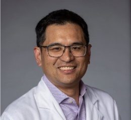
Duke University Medical Center
Title: Machine Learning Algorithm Enabled Thyroid Nodule Cytopathology : Detecting Cancer in Resource Limited Settings
Dr. Walter T. Lee MD MHS is Professor in the Duke University Department of Head and Neck Surgery & Communication Sciences He currently serves as the Department Chief of Staff and Co-Director of the Head and Neck Program in the Duke Cancer Institute. He has joint appointment at the Duke – National University of Singapore and also is an affiliate faculty member at the Duke Global Health Institute.
His research interests are focused on two areas. The first is developing technology and approaches to improve the diagnosis and treatment of head and neck cancers in patients living in low resource settings. This has included device development and nanotechnology for early cancer detection. He holds several NIH funded grants that currently work with collaborators in Vietnam, Tanzania, and Singapore, The second is a focus on professional formation and leadership development. He was co-creator of a virtue-based approach to leadership and professional development.
Objectives
This study aims to prospectively implement and validate a machine learning algorithm (MLA) for analysis of smartphone captured fine needle aspiration biopsy (FNAB) images of thyroid nodules in tertiary centers in Tanzania and Vietnam, correlating MLA assessment with cytopathology to determine diagnostic performance in low and middle income country (LMIC) settings.
Scope
Images were collected at Kilimanjaro Christian Medical Center (Tanzania) and the National Otolaryngology Hospital (Vietnam) using Redmi Note 10S smartphone cameras mounted on optical microscopes. A total of 3,167 smartphone captured ROI images were obtained: 1,119 images from 100 patients in Tanzania and 2,048 images from 132 patients in Vietnam. One non-pathologist at each site was trained by the local pathologist to identify and photograph regions of interest.
Methods Used
Fine-tuning a MobileNet-v2 model, originally trained on Duke data, on the Tanzania and Vietnam datasets produced an approximate 10% drop in accuracy, attributed to differences in staining and imaging equipment across sites. Site-specific EfficientNet-b0 models were then trained directly on each dataset using surgical histopathology diagnosis as the ground truth label. Predictions from multiple images per patient were combined by majority voting, and explainable AI heatmaps were generated to highlight regions driving each prediction.
Results
On the Tanzania test set (20 patients), the model achieved 90% accuracy, 91% F1-score, 100% sensitivity, and 89% specificity. On the Vietnam test set (40 patients), it achieved 95% accuracy, 95% F1-score, 97% sensitivity, and 89% specificity. Heatmaps largely localized to diagnostically relevant cellular regions, supporting model explainability.
Conclusion
Site-specific model training overcame image heterogeneity between LMIC sites, yielding strong diagnostic performance, while demonstrating that non-pathologists can be trained to capture adequate smartphone images for AI-assisted thyroid cytology in resource-limited settings.
